Vascular Malformations of Spine
Spinal cord vascular malformations are abnormal connections between arteries and veins in the spinal
column due to the absence of small blood vessels or capillaries. This causes the abnormal flow of blood.
Spinal vascular malformations include the malformations of the spinal cord, vertebral bodies, the tissue
around the spinal cord & the spine, the bones of the spine, or a combination of these. They are serious
medical issues that can even result in paralysis if left untreated
The disorders include:
- Spinal arteriovenous malformations (AVMs)
- Dural arteriovenous fistulas (DAVF)
- Spinal hemangiomas
- Cavernous angiomas
- Cavernous malformations
- Aneurysms
Cause of Vascular malformations
Although the specific cause of these congenital abnormalities is not known, the following reasons may cause vascular malformations:
- Congenital abnormalities of blood vessels occur in the younger patient population
- Subarachnoid hemorrhage
- Increased venous pressure
- Progressive damage to the spinal cord
- Trauma in the older population
Symptoms of Vascular malformations
Some of the spinal cord vascular malformations are congenital while some develop later in life. They show the following symptoms either acutely or may take several years to appear:
- Either loss of sensations or abnormal sensations such as numbness or tingling
- Sudden onset of pain in back, neck, or leg
- Extreme weakness in the upper or lower extremity
- Loss of bladder and bowel control
- Progressive neurological deficits
- Headache
- Neck stiffness
- Photosensitivity or intolerance of bright light
- Paralysis
- Pain radiating down the legs
Diagnosis of Vascular malformations
The diagnosis of malformations requires a complete history of the patient and a thorough physical and neurological examination. This along with following diagnostic tests helps the physician to appropriately treat the abnormality:
- MRI
- Myelography
- Spinal angiography
Treatments
Medication:
Pain-relieving medications may be used to reduce symptoms such as back pain and stiffness but most spinal AVMs eventually require surgery.
Surgery:
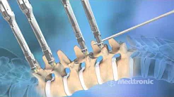
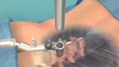
Conventional surgery: In this procedure, a surgeon makes an incision to remove the AVM taking care to avoid damaging the spinal cord and other surrounding areas. Surgery is usually performed when the AVM is fairly small and in an area of the spinal cord that's easy to reach out.
Endovascular
embolization:
Endovascular embolization is a minimally invasive radiological procedure used to reduce the risk of hemorrhage and other complications associated with spinal AVMs.
In endovascular embolization, a catheter is inserted into an artery in your leg and threaded to an artery in your spinal cord that are feeding your AVM. Small particles of a glue like substance are injected to block the artery and reduce blood flow into the AVM. It does not permanently destroy the AVM.
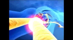
Radiosurgery: such procedure uses radiation focused directly
on the AVM to destroy the blood vessels of the malformation. Over this time,
these blood vessels break down and close up. Radiosurgery is most often used to
treat small, unruptured AVMs.


 Request an Appointment
 Request Online Consultation
 Enquiry form


